[BioSB] TintoFast EpCAM BerEP4 (Ber-EP4), MMab
| Intended Use | For Mohs In Vitro Diagnostic Use | |||
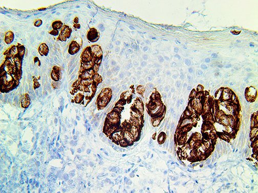
| Summary and Explanation | TintoFast EpCAM BerEP4 is a 40kD cell surface antigen that is broadly distributed in epithelial cells and displays a highly conserved expression in carcinomas. These glycoproteins are located on the cell membrane surface and in the cytoplasm of virtually all epithelial cells, with the exception of most squamous epithelia, hepatocytes, renal proximal tubular cells, gastric parietal cells and myoepithelial cells. However, focal positivity may be seen in the basal layer of squamous cell epithelium of endoderm (e.g., palatine tonsils) and mesoderm (e.g., uterine cervix). TintoFast EpCAM BerEP4 expression has been reported to be a possible marker of early malignancy, with expression being increased in tumor cells, and de novo expression being seen in dysplastic squamous epithelium. Epithelial specific antigen has been known to play an important role as a tumor-cell marker in lymph nodes from patients with esophageal carcinoma. EpCAM can be used to distinguish among Basal Cell, Basosquamous Carcinomas and Squamous Cell Carcinomas of the skin. |
|||
| Antibody Type | Mouse Monoclonal | Clone | Ber-EP4 |
| sotype | IgG1/K | Reactivity | Paraffin, Frozen |
| Localization | Cytoplasmic | Control | BCC, EMPD |
| Presentation | Anti – EpCAM BerEP4 is a mouse monoclonal antibody derived from cell culture supernatant that is concentrated, dialyzed, filter sterilized and diluted in buffer pH 7.5, containing BSA and sodium azide as a preservative. | |||
| Catalog No. | Antibody Type | Dilution | Volume/QTY |
| BSB 3678 | TintoFast Prediluted | Ready-To-Use | 3.0 ml |
| BSB 3679 | TintoFast Prediluted | Ready-To-Use | 7.0 ml |
| BSB 3680 | TintoFast Prediluted | Ready-To-Use | 15.0 ml |
Mohs IHC Procedure
Specimen Preparation of Mohs Frozen Tissues
- Embed the specimen in OCT inside a cryostat.
- Cut sections at 4-5 µm and mount on a positively charged glass slide such as the Bio SB Hydrophilic Plus Slides (BSB 7028) or in the lower third of the TintoDetector Cap Gap slides (BSB 7006).
- Air dry the slide at room temperature for 2 minutes and then incubate the slide at 60 °C for 3 minutes in an incubator or dry bath.
- Fix in 100% acetone for 2 minutes at room temperature.
- Rinse with distilled water and air dry the slides for another 2 minutes at room temperature.
IHC Detection Procedure
- Transfer slides to ImmunoDNA washer, TBST or PBST buffer.
- For manual staining, perform antibody incubation at ambient temperature. For automated staining methods, perform antibody incubation according to instrument manufacturer’s instructions.
- Wash slides with ImmunoDNA washer, TBST, PBST or DI water.
- Continue IHC detection protocol. Wash slides between each step with ImmunoDNA washer, Tris or PBS Buffer solution.Abbreviated Mohs Immunohistochemical Protocol
-
- Do HIER with Citrate in Pressure Cooker (100 – 121 °C) for 5 min. Cool off
- Wash with Buffer (TBST or PBST)
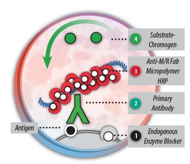
- Incubate slides with Peroxidase Blocker for 30 seconds
- Wash with Buffer (TBST or PBST)
- Incubate Primary Antibody for 4 min
- Wash with Buffer (TBST or PBST)
- Incubate with HRP Label for 3 min.
- Wash with Buffer (TBST or PBST)
- Prepare
- DAB Brown (1 drop of DAB chromogen in 1 ml of DAB Buffer; mix well)
- or HRP Green (1 drop of HRP Green chromogen in 1 ml of HRP Green Buffer, mix well)
- Incubate with DAB or HRP Green for 2 min
- Wash with Buffer (TBST or PBST)
- Counterstain with Hematoxylin or Nuclear Fast Red for 30 seconds
- Wash with Buffer (TBST or PBST)
- Mount with AquaMounter or PermaMounter
For best results we recommend using Mohs Frozen sections fixed NBF10% for 2 min., then HIER with Citrate for 5 min. at 110 – 121 °C.
Step Mohs PolyDetector HRP Green 5 min Protocol Mohs PolyDetector HRP Green 10 min Protocol Peroxidase Blocker 0.5 min. 0.5 min. Primary Antibody 2 min 4 min. 1st Step Detection 1 min 3 min. Substrate-Chromogen 1 min 2 min. Counterstain / Coverslip 0.5 min 0.5 min. 
This protocol can also be used with FFPE Tissues retrieved with Citrate or EDTA.
'BioSB' 카테고리의 다른 글
| [BioSB] 11 Core Normal Human Tissue Microarray (0) | 2024.02.27 |
|---|---|
| [BioSB] TintoFast Cytokeratin 5 & 6 (D5/16D4), MMab (0) | 2023.05.08 |
| [BioSB] 23 Core Normal Human Tissue Microarray (0) | 2021.05.26 |
| [BioSB] ZytoLight FlexISH Probes and Kits (0) | 2021.05.17 |
| [BioSB] TintoFast CD34 – MMab (0) | 2021.02.03 |


